
Illustration of the gastric vessels and the ligatures of their... Download Scientific Diagram
Dr. O'Boyle and associates have published 5-year follow-up data from their prospective randomized trial of laparoscopic Nissen fundoplications performed with and without division of the short gastric vessels. 1 Early results of this trial, which suggested no difference between the two groups other than an increased operating time in those underg.

The Spleen Basicmedical Key
Kinsey-Trotman et al. followed 102 patients randomized to either division or non-division of short gastric vessels during laparoscopic Nissen fundoplication for 15-20 years, and found that fewer patients who did not have division of the short gastric vessels report heartburn (RR = 0.73, 95%CI = 0.46, 1.14, high risk of bias). 20 Mardani et al.
Where Are Short Gastric Arteries From
The technique of coagulating the peripheral gastric vessels to prevent SLB is safe and appears promising. A prospective study comparing with and without peripheral gastric vessel coagulation will be needed in the future. Keywords: Bariatric surgery, bleeding, complication, sleeve gastrectomy, staple line Go to:
Stomach Anatomy Earth's Lab
This retrospective study is based on short gastric vessels division (SGVsD), if necessary, during LF to construct a satisfactory loose wrap and to evaluate its effect upon the symptomatic and physiologic outcome in patients with proven GERD.
Short gastric arteries Alchetron, The Free Social Encyclopedia
Releasing the short gastric vessels is performed first because the remaining attached pedicle is long and the entire spleen can be easily laid outside the abdomen for pedicle isolation after this maneuver. The figure shows the short gastric pedicle incised between forceps #2 and 3, freeing the tethered head of the spleen.

Note 3 Branches of Celiac trunk / artery imp. 【 Common hepatic artery Gastroduodenal
Figure 18-1: The gastrosplenic ligament contains the short gastric vessels and must be divided to obtain access to the splenic vessels, whereas the splenophrenic and splenorenal ligaments are relatively avascular.; Figure 18-2A, B: Laparoscopic splenectomy can be performed either in the supine (Figure 18-2A) or right lateral decubitus (Figure 18-2B) positions using similar port placement.
Arteries Adjacent to Stomach (Labeled) Eccles Health Sciences Library J. Willard Marriott
These include whether to perform partial versus total fundoplication, divide the short gastric vessels or not, whether to use a laparoscopic or robotic approach, or to provide minimal or maximal dissection in the pediatric population. These guidelines provide recommendations regarding the surgical treatment of GERD.
What Does The Short Gastric Artery Supply
In hypotensive patients, the short gastric vessels usually do not bleed, nor does the splenic bed. In the case of elective splenectomy, the first step is transection of the ligamentous.
PPT Anatomy of stomach and its relations PowerPoint Presentation, free download ID9343361
Methods: Literature reviews were conducted for 4 key questions regarding the surgical treatment of GERD in both adults and children: surgical vs. medical treatment, robotic vs. laparoscopic fundoplication, partial vs. complete fundoplication, and division vs. preservation of short gastric vessels in adults or maximal versus minimal dissection in.
The right gastric artery Anatomy, branches, supply Kenhub
Dividing the short gastric vessels during surgery for gastroesophageal reflux is controversial. This prospective randomized study was designed to determine whether there is a benefit in terms of patient outcome at a minimum of 5 years after primary surgery. Methods

Right gastric artery
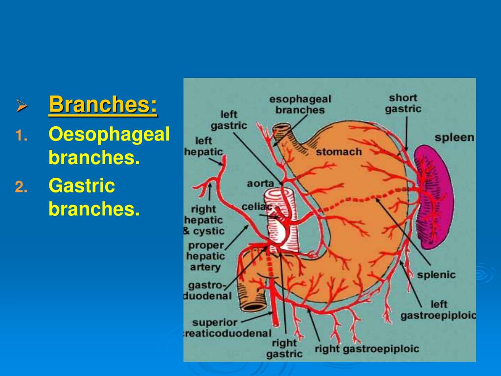
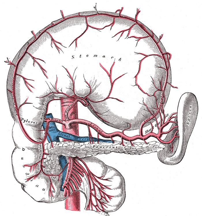
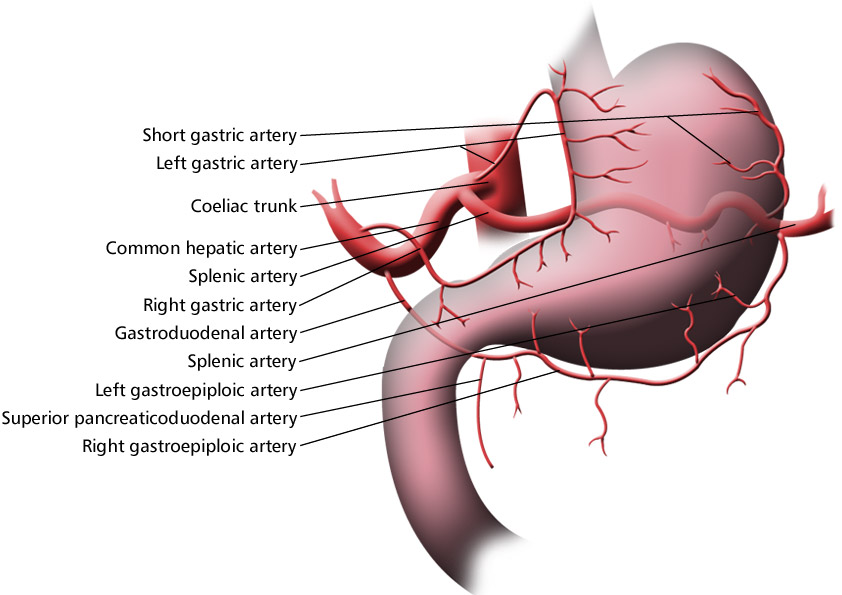
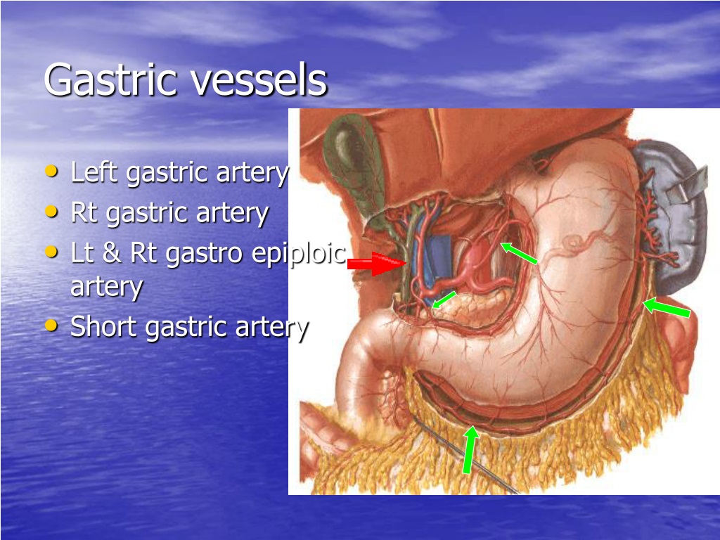
The short gastric arteries are a group of short arteries arising from the terminal splenic artery and the left gastroepiploic artery which supply the fundus of the stomach along its greater curvature. The vessels are short in length, variable in number and course through the gastrosplenic ligament to the gastric fundus.
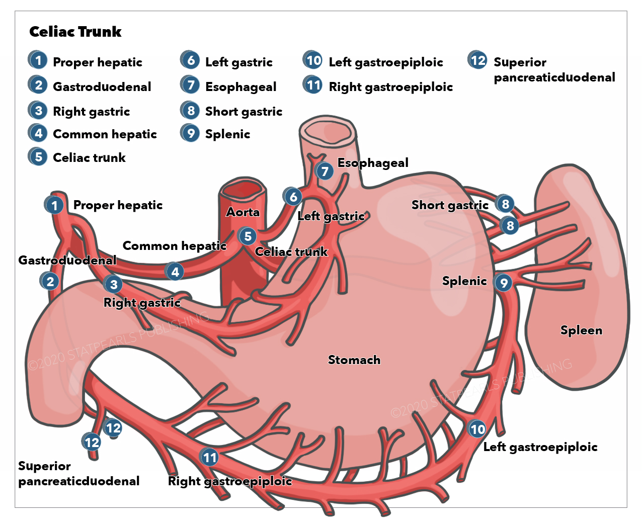
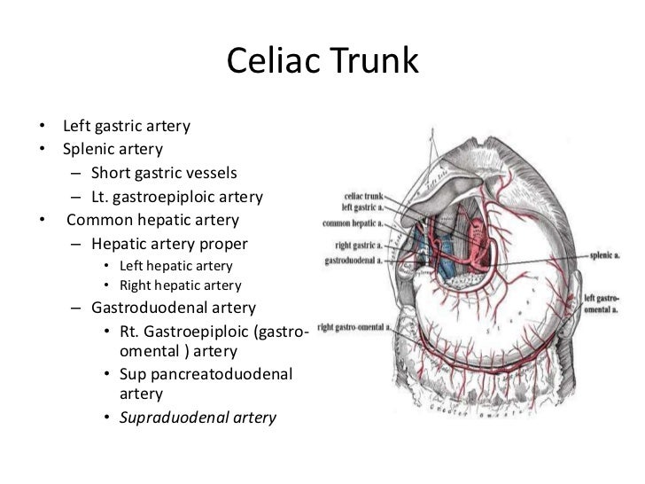
Anatomy, Abdomen and Pelvis, Celiac Trunk Article
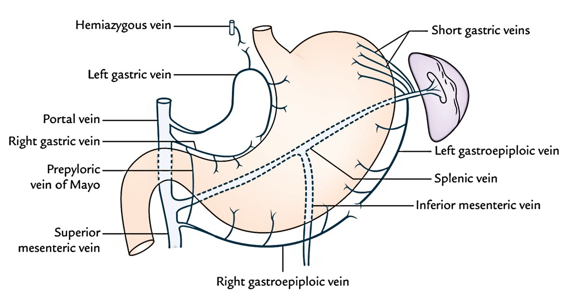
The veins of the gastric cardia and gastric fundus drain into the coronary vein in the lesser omentum, the short gastric veins in the gastrosplenic ligament, and paraesophageal and esophageal veins. Any condition that elevates pressure in the splenic vein can result in splenic blood draining through the short gastric vessels, retrogradely into.

Ligation of the short gastric vessels for mobilization of the gastric... Download Scientific
The gastrosplenic ligament (also known as the ligamentum gastrosplenicum or gastrolienal ligament) is part of the greater omentum extending between the stomach and the spleen. It contains several blood vessels. Structure The gastrosplenic ligament consists of visceral peritoneum. [1]

Celiac Trunk Gastrointestinal Medbullets Step 1
The short gastric veins are located on the left side of the stomach, between the gastrolienal ligaments. The veins act as a drainage system for the stomach, particularly around the fundus..

Stomach and esophagus The esophagus is a tubular
The stomach is an organ of the digestive system, specialized in the accumulation and digestion of food. Its anatomy is quite complex; it consists of four parts, two curvatures and receives its blood supply mainly from the celiac trunk. Innervation is provided via the vagus nerves and the celiac plexus .

Pathogenesis, Diagnosis, and Management of Gastric Ischemia Clinical Gastroenterology and
The splenic artery also gives off 3-5 short gastric arteries that run in the gastro-splenic (gastro-lienal) ligament and supply the upper part of the greater curvature and the gastric fundus. Few small posterior gastric arteries may arise from the splenic artery. The stomach has a rich network of vessels in its submucosa.